New & Noteworthy
Mass Production in Yeast
May 11, 2017

This approach changed everything when it came to manufacturing in factories. Perhaps the ideas in this new study will change things for manufacturing in cells.Image from Wikimedia Commons
After Henry Ford invented the moving assembly line, manufacturing was never the same. With it, his workers were able to push out a car every 2 ½ hours instead of the 12 it used to take. (Another website said it was reduced to 90 min!) The technology quickly spread to every factory.
Now of course, an assembly line is only as fast as its slowest worker. If someone is taking extra time to bolt down that part, then everyone downstream will have to go slower too, resulting in fewer cars being made.
But, you also can’t go too fast. If you do, someone can get injured, shutting down the whole line. (Or the worker has to eat all the candy to keep up, like Lucy.)
And you want to make sure things happen in the right part of the factory. You don’t want the paint sprayer out in the open, poisoning factory workers. So, that needs to happen in a special room.
This also applies to cell processes where something complicated is built, step by enzymatic step. All the enzymes need to be at the right levels and in the right place to maximize the productivity of the whole process.
This all becomes very obvious when you try to move an enzymatic process from one beast to another. What worked perfectly before, now barely works at all.
One way to fix this is through trial and error, trying to optimize one part of the process at a time. This is incredibly time consuming!
In a new study out in Nature Communications, Awan and coworkers show one way to tweak all of the enzymatic steps involved in making penicillin at the same time in the yeast Saccharomyces cerevisiae. While this isn’t that useful for making this antibiotic (there are better ways available right now), it does show how researchers can apply the same techniques to perhaps identify and produce new antibiotics. And, it can also be applied to other unrelated enzymatic processes.
Penicillin is made in a five-step process in filamentous fungi. In the first part of the process, two enzymes create a tripeptide precursor using alpha-aminoadipic acid, cysteine, and valine, called ACV. This part of the process had been previously recapitulated in yeast, so Awan and coworkers used this as a starting point for their penicillin producing strain.
The next part of the process uses the last three enzymes and takes place in peroxisomes in filamentous fungi. These authors found that they only got penicillin when these enzymes were tagged to be sent to the peroxisome in yeast. Like a special room for spray painting cars, these enzymes need to be in the right place to make penicillin.
But this was by no stretch of the imagination an efficient penicillin-making machine. The thing managed only 90 pg/ml in the media. As Ursula from Little Mermaid might say, “Pathetic.”

From Tumblr
Still, it is a starting point. The next step is to get the yeast to crank out more penicillin. To do this, they used a combinatorial approach to optimize the process all at once. Well, not really all at once.
First, they set out to optimize how much of the precursor ACV the yeast made. Then, they optimized how much ACV was converted to penicillin.
Awan and coworkers created a library of low copy plasmids that had the genes for the first two enzymes, pcbAB and npgA, under the control of different pairs of promoters. One plasmid, with the pTDH3 promoter driving pcbAB expression, and the pPGK1 promoter driving npgA expression, outperformed all of the others. As measured by Liquid Chromatography-Mass Spectrometry (LCMS), the yield of ACV increased from 20 to ~280 ng/ml.
Next, the authors used this new strain as a starting point for optimizing the activity of the final three enzymes using a similar approach. They used a “…one-pot combinatorial DNA assembly using Golden Gate cloning…” to make a library of around 1000 high copy plasmids where each gene was under the control of one of ten different promoters of varying strength. Using LCMS they found strains that could make 3 ng/ml of penicillin, a significant improvement over the original 90 pg/ml.
The 3 ng/ml of penicillin in the media should be high enough concentration to inhibit the growth of bacteria like Streptococcus pyogenes. So, they confirmed that their penicillin was active using growth inhibition assays.
After sequencing the plasmids, the authors saw that the best strains tended to have strong constitutive promoters driving one of the genes, pclA, and medium strength promoters driving another one of the genes, pcbC. They used a minION DNA sequencer to confirm that this was not the result of a biased library.
As a final step, they set out to optimize penicillin production and to increase the throughput of their assay. They created another library that swapped six different promoters that varied in strength from medium to high for each of the last three genes in the pathway, pclA, pcbC and penDE. Instead of using LCMS to screen for penicillin production, they used a 96 well plate-based assay that looked for inhibition of Streptococcus pyogenes growth for their 120 new strains.
They selected 12 of the highest performing strains and confirmed by LCMS that they made lots of penicillin. Five of the strains made more than 5 ng/ml, a more than 50-fold increase over their original strain.
As this concentration is still three orders of magnitude below what other organisms can currently do, this new yeast strain will not go into penicillin production any time soon. But this study gives us a way to quickly optimize antibiotic production using growth inhibition assays instead of the more cumbersome LCMS.
And it isn’t restricted to just antibiotic production. Similar combinatorial approaches can be used for almost any stepwise enzymatic process. Researchers can create libraries of plasmids where levels of enzyme vary and use the long reads of minION DNA sequencing technology to confirm that their results are not skewed by a biased library.
As usual, this is only possible as a simple, easy procedure because of the awesome power of yeast genetics (#APOYG). Researchers have the tools to use yeast to find new antibiotics and to manufacture them at a high rate, like inventing the car and the assembly line at the same time.
by Barry Starr, Ph.D., Director of Outreach Activities, Stanford Genetics
Categories: Research Spotlight
Tags: ACV, liquid chromatography-mass spectrometry, minION, npgA, pcbAB, pcbC, pclA, penDE, penicillin synthesis
Meiotic Fail Safes
May 04, 2017
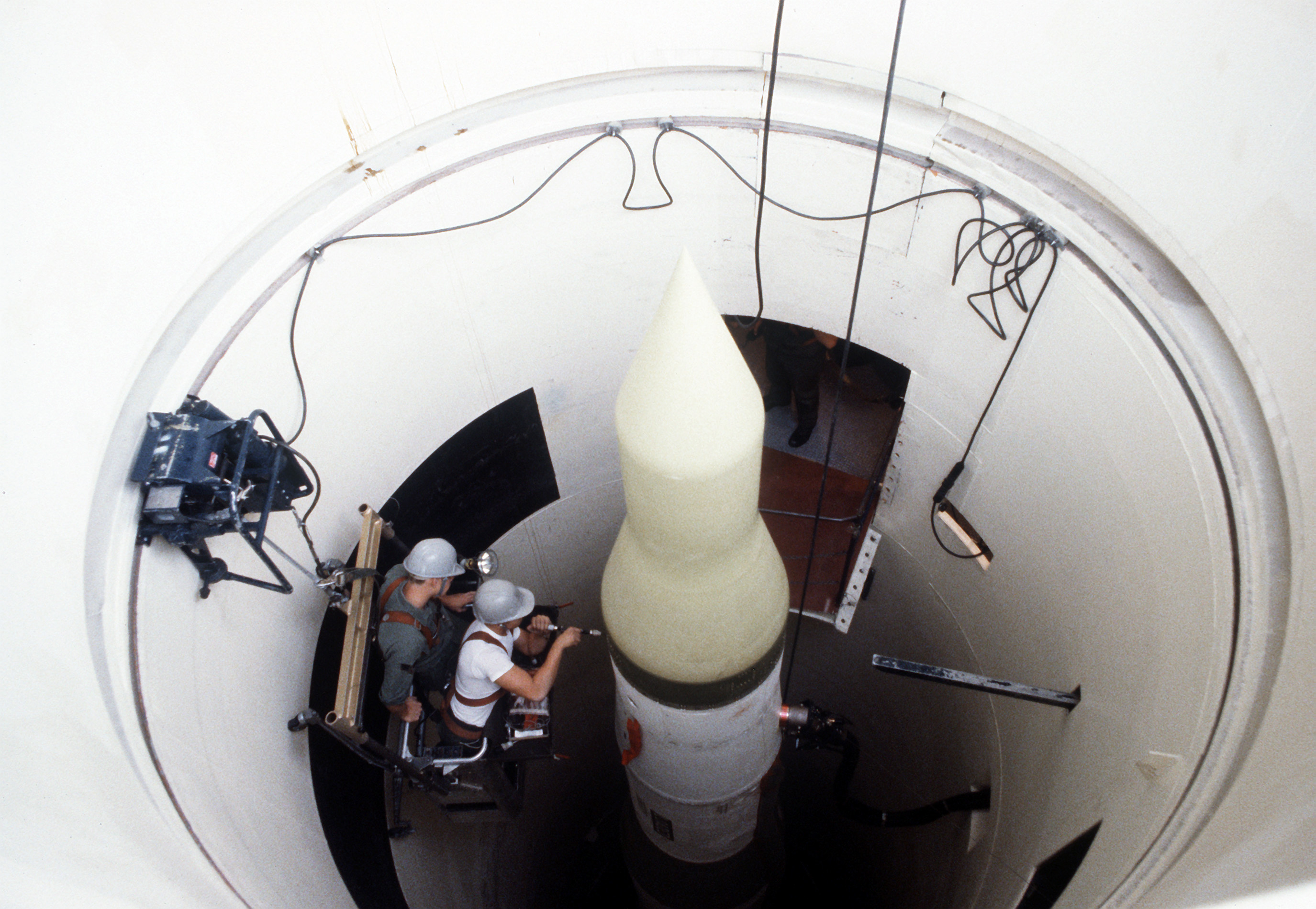
Launching into meiosis too soon is as dangerous (for a cell) as launching a nuclear missile. Luckily both have protocols to make sure each can only happen in the right circumstances. (Hopefully never for the nuclear missile.) Image from Wikimedia Commons.
If the movie WarGames is anything to go on, the US government does not make it easy to launch a nuclear missile. Two soldiers have to do many things simultaneously and in the right order before that missile can take flight.
This makes perfect sense as you do not want to launch a nuclear attack unless you absolutely have to. The continued existence of the human race depends on these fail safes being in place and working. The same goes for a cell that is heading into meiosis.
Meiotic fail safes are in place to ensure the survival of a cell during the dangerous, early part of meiosis, when there are lots of double-strand breaks in the DNA. These all need to be resolved before a cell is allowed to continue through meiosis to create gametes. If the cell moves on while the breaks are still there, gamete production will fail and the cells will die.
While the exact sequence of events needed to launch World War III is known (at least by a few people), the exact details of getting a cell safely through meiosis are a bit murkier. With the help of good old Saccharomyces cerevisiae, we have the broad outlines, but are still investigating the finer points.
A new study by Prugar and coworkers in GENETICS has helped clear up a bit of the murk in yeast. They have uncovered a connection between the meiosis-specific kinase Mek1p and the transcription factor Ndt80p that may explain how a cell “knows” when it is safe enough to emerge from prophase and keep progressing through Meiosis I.
Mek1p is known to be active when there are lots of these double-strand breaks around and to lose activity as these breaks are resolved. Ndt80p, on the other hand, is inactive when there are lots of these breaks and active when they are resolved. So it makes sense that their activities might be related to each other.
In this study, the authors show that once Mek1p activity falls below a certain level, it can no longer keep tamping down Ndt80p activity. Once unleashed, Ndt80p can go on to activate many genes, including the polo-like kinase CDC5 and the cyclin CLB1. This round of gene activation allows the cell to progress through meiosis.
The key to teasing this out was a set of experiments where Prugar and coworkers were able to control the activities of Mek1p and Ndt80p independent of the cell’s DNA state. It is like circumventing the set of protocols to get those missiles launched.
To independently control Ndt80p activity, they used a form of the protein that requires estradiol to be active. And they controlled the activity of Mek1p by using a mutant, mek1-as, that is sensitive to the purine analogue 1-NA-PP1. In the presence of this inhibitor, Mek1p stops working.
They looked at the targets of these two proteins to infer activity. For example, they determined if Ndt80p was active by looking for the presence of CDC5. And to see if Mek1p was active, they looked for phosphorylated Hed1p.
In the first experiment, they showed that in the absence of both estradiol and 1-NA-PP1, Hed1p stayed phosphorylated. Mek1p was constitutively active in the absence of Ndt80p even as double-strand breaks were resolved. (They used phosphorylated Hop1p as an indirect measure of double-strand breaks.)

In the end, as yeast relies on MEK1 to prevent a meiotic disaster, humans, not computers, kept the world safe in the movie WarGames. Image from flickr
When Ndt80p was activated through the addition of estradiol, CDC5 was turned on and Hed1p lost its phosphorylation. This loss of Mek1p activity did not happen as quickly as with 1-NA-PP1.
These results suggest a negative interaction between Mek1p and Ndt80p. When Ndt80p is active, Mek1p is not and when Mek1p is active, Ndt80p is not. The resolution of the DNA breaks as indicated by the loss of phosphorylated Hop1p was not sufficient to shut off Mek1p activity. It took the activation of Ndt80p for this to happen.
Well, Ndt80p did not directly cause Mek1p’s inhibition. A second set of experiments suggested that a target of Ndt80p, CDC5, was responsible.
For this they made Cdc5p activity independent of Ndt80p induction by making it dependent on estradiol, similar to what they did with Ndt80p. Using a strain deleted for NDT80, they found that inducing Cdc5p activity was enough to eliminate Mek1p activity.
I don’t have the space to go into the rest of the experiments in this study, but I urge you to read it if you want to learn about more of the details of the cell’s protocol for know when it is OK to progress through meiosis.
With the help of the awesome power of yeast genetics (#APOYG), Prugar and coworkers have added to our knowledge about the safeguards that are in place to keep a cell from launching into meiosis too soon. Turns out they are even more complicated than the ones that prevent accidental thermonuclear war.
by Barry Starr, Ph.D., Director of Outreach Activities, Stanford Genetics
Categories: Research Spotlight
Tags: CDC5, double-strand breaks, DSB, meiosis, MEK1, NDT80
Knock out YME1, Luke
April 20, 2017

Instead of an exhaust port, one of a mitochondrion’s fatal flaws may be the YME1 gene. Image from Manoel Lemos, flickr.
In the original Star Wars, Luke destroys the Death Star with a precise strike of proton torpedoes down a small thermal exhaust port. For him it was as easy as bullseyeing “womp rats in my T-16 back home.”
Luke and the rest of the Rebel Alliance learned of this engineered fatal flaw from Jyn and her friends in the prequel Rogue One. With this information the Rebel Alliance was able to keep the rebellion alive long enough to finally bring down the Empire by the end of Return of the Jedi.
It turns out that our friend Saccharomyces cerevisiae has taught us about a fatal flaw in mitochondria. Like proton torpedoes in an exhaust port, when the gene YME1 is inactivated, mitochondria become unstable. But instead of bits of Death Star raining down on nearby planets, mitochondrial DNA (mtDNA) is released into the cytoplasm.
Sometimes this mtDNA can end up in the nucleus and find its way into nuclear DNA. And if the conclusions of a new study in Genome Medicine by Srinivasainagendra and coworkers turns out to be right, this numtogenesis (as the authors call this process) can have profound consequences when it happens in people. Their data suggests that it might lead to cancer or possibly cause cancers to spread.
These researchers searched through whole genomes of colon adenocarcinoma patients and found that these cancer cells had 4.2-fold more mtDNA insertions compared to noncancerous cells from the same patient. They also found that patients with more of these insertions tended to do worse (although the sample sizes were too small to say this definitively).
Why is this happening in the cancer cells? What has caused the mitochondria to give up their DNA?
Srinivasainagendra and coworkers turned to previous work that had been done on the YME1 gene in the yeast S. cerevisiae to find one possible reason. YME1 had been shown to be an important suppressor mtDNA migration to the nucleus. Perhaps this was true in mammalian cells as well.
A search through the genomes of cancers suggested that this seemed to be the case. Around 16% of the colorectal tumors they looked at had a mutated YME1L1 gene, the human homologue of YME1. And mutated YME1L1 genes were found in other tumors as well.
If only destroying gene function was as fun.
They used CRISPR/Cas9 to directly test the effects of knocking out YME1L1 in the breast cancer cell line MCF-7. The knock out cells had a 4-fold increase in the amount mtDNA in the nuclear fraction compared to cells that still had working YME1L1.
As a final experiment, they used a yeast strain, yme1-1, in which YME1 function was inactivated, to show that the human homologue, YME1L1, could suppress the migration of mtDNA to the nucleus.
This yme1-1 strain has a TRP1 gene encoded in the mtDNA instead of the nucleus. Since the gene cannot be read by the mitochondrial transcription machinery, the only way this yeast strain can survive in the absence of tryptophan is if the TRP1 gene moves from the mitochondrion to the nucleus.
In their experiment, with vector alone, they got around 1000 TRP+ colonies with yme1-1. When they added back yeast YME1, this number dropped to less than 50 compared to the 100 or so they got when they added the human homologue, YME1L1. So YME1L1 can suppress mtDNA migration to the nucleus.
Given that YME1L1 was mutated in just a subset of the cancers, it is unlikely that it is the only player in the mtDNA these authors found in the nuclei of cancer cells. But it does look like it is one way this can happen.
And it would have been very hard to fish out the human gene without the critical work that had been done in yeast previously. Yeast shows us the way again. #APOYG
by Barry Starr, Ph.D., Director of Outreach Activities, Stanford Genetics
Categories: Research Spotlight
Tags: cancer, colon cancer, CRISPR/Cas9, MCF-7, mtDNA, nuMt, nuMtogenesis, YME1, YME1L1
The Dark Yeast Rises
April 04, 2017

Sometimes in life you need to take risks to survive and prosper. Maybe you need to take a leap of faith like Bruce Wayne does to escape that pit in The Dark Knight Rises. Or you need to try a risky business strategy to put your company out in front.
While these are critical things to do at the time, chronic risk-taking is not usually a good idea. Even Bruce Wayne retires to Florence at the end of The Dark Knight Rises, his risky life choices done now that he has saved his beloved Gotham City. He can now settle down with Selina Kyle (Catwoman) and live happily (and safely) ever after.
Something similar can happen in the budding yeast, Saccharomyces cerevisiae. In the right environment, certain yeasts can build up mutations like mad, hoping to hit on one that lets them make it out of that pit.
But then, over time, yeasts with the successful mutation lose that frenetic mutation rate—they take fewer risks with their DNA. One way they can do this is by ending up with a mutation that suppresses the high mutation rate. This is like Bruce Wayne retiring to a nice villa on the Arno.
Another way they can reestablish their old mutation rate is to mate with a nonmutator, a yeast strain that does not risk its DNA with a high mutation rate. Now, the next generations have the beneficial mutation and a lowered mutation rate as well. It is as if Bruce Wayne settled down with an accountant instead of Catwoman and had risk-averse children to carry on the Wayne name.
Bui and coworkers investigate this phenomenon in yeast in a new study out in GENETICS. They show that natural isolates exist that can sporulate into one of these mutator strains. And that at least one strain predicted to have a high mutation rate has a number of suppressor mutations that tamp down that higher rate.
Previous work had shown that a high mutation rate can happen with mutations in two genes in the mismatch repair (MMR) pathway, MLH1 and PMS1. This was discovered when researchers saw that some of the haploid strains from a mating of two laboratory strains, S288C and SK1, showed this mutator phenotype. A closer look revealed that S288C has a mutation in MLH1, and SK1 has a mutation in PMS1.
Bui and coworkers show that these mutator haploid strains outcompete other strains in high salt media. The strain hits upon mutations that allowed for better survival in high salt faster than its slower mutating brethren. But this advantage does not last.
After 120 generations these mutator strains start to lag behind strains with more typical mutation rates. Their DNA becomes as beat up as Bruce Wayne’s body by the third movie.
The researchers next looked at the MLH1 and PMS1 genes of 1,010 natural isolates of S. cerevisiae to see if there were any that might be able to sporulate into a mutator strain. Or that were already mutator strains themselves.
Since the mutations that lead to the mutator phenotype are recessive, strains that are heterozygotes for mutations in both genes should be able to form haploid spores with the higher mutation rate. They found 18 that fit the bill.
They also found one strain, YJM523, that was homozygous for both mutations and so would be predicted to have a high mutation rate. They further investigated this strain.

The first thing they did was to test the YJM523 mutant genes in an S288C background. They found that these two mutant genes did indeed give S288C a mutator phenotype. In fact, it had a higher mutation rate than the original genes from S288C and SK1 did, suggesting there were other mutations in one or both YJM523 genes that further increased the mutation rate.
These researchers next wanted to determine if YJM523 had a higher mutation rate or not, but needed to first develop a new assay that could be used in natural isolates. They came up with a reporter system that is not dependent on the typical markers used in yeast experiments, but instead relies on two antibiotic markers.
The reporter uses the NatMX antibiotic resistance marker for selection and the KanMX marker to identify revertant mutants. By scoring the number of colonies resistant to both antibiotics, they could determine the mutation rate.
When the researchers did this experiment, they found that YJM523 did not have a particularly high mutation rate. Sporulation experiments showed that this was most likely due to multiple suppressor mutations.
So out in the wild, the potential for mutator strains exists. And a close look at the genome of YJM523 showed that if it did have a mutator phenotype, it was for a short time. This yeast at least quickly lost its high mutation rate (assuming it ever had one).
This study helps to explain how yeast can obtain that mutator phenotype that researchers have seen before. And how yeast can escape that mutation pit to get back to the safety of a reasonable DNA repair pathway. To quote the prisoners in The Dark Knight, “Deshi Basara”, or ‘rise up’ yeast!
by Barry Starr, Ph.D., Director of Outreach Activities, Stanford Genetics
Categories: Research Spotlight
Tags: adaptation, DNA mismatch repair, experimental evolution, genetic incompatibility, mutator phenotype, natural yeast isolates
Too Big for the Oven
March 23, 2017

Just like baking bread, “oven” size matters when trying to increase yields of fatty acids in yeast.
In a classic skit from I Love Lucy, Lucy bakes a loaf of bread that is so big it ends up coming out of the oven, pushing her against the far wall. Definitely one of the funniest moments from early TV.
In a new study in Nature Communications, Gajewski and coworkers show that the yeast enzyme fatty acid synthase (FAS) complex, is a bit less comical when it comes to the fatty acid chains it makes. When these authors engineer the enzyme complex to “shrink” the oven, the “bread” it makes becomes smaller. A more constrained active site that holds onto its growing fatty acids less tightly makes shorter-chained fatty acids.
This is an important finding because these shorter-chained fatty acids are so useful as precursors for products like biofuels. Engineered yeast like these may, among other things, one day help us get to a carbon neutral future.
Gajewski and coworkers were able to pull this off because the 3-D structure of this enzyme complex is so well understood. They engineered changes that would be predicted to restrict the growth of the fatty acid chain.
For example, a key player in the elongation step happens in the condensation domain (KD) of the complex. They focused initially on the amino acid methionine at position 1251 (M1251).
This amino acid sticks out into the active site and needs to rotate out of the way when the fatty acid chain elongates. The authors reasoned that if they made it bigger and harder to rotate, the enzyme complex wouldn’t be as good at elongating the fatty acid chains it makes. The loaf would stop growing when it ran out of room.
They were right. When they mutated a nearby glycine to serine (G1250S), the yeast now made 15.3 mg/liter of fatty acids with 6 carbons (C6 fatty acids), a 9-fold increase over wild type. FAS usually makes C12-C18 fatty acids.
Tomorrow’s oil wells? By (Image: Maitreya Dunham) [CC BY 2.5], via Wikimedia Commons
They got even better results when they bulked up position 1251 by changing the methionine to a tryptophan (M1251W). Now the yeast made 19.1 mg/liter of C6 fatty acids, a 12-fold increase, and 32.7 mg/liter of C8 fatty acids, a 56-fold increase. They made a third mutation, F1279Y, that ended up affecting growth adversely when combined with G1250S.
This was good, but not good enough. To be commercially viable, they needed higher yields. They next wanted to make mutations that would cause the enzyme complex to hold onto the fatty acids less tightly, like having the baker remove the bread from the oven before it got too big.
For this, they focused on the malonyl/palmitoyl transferase (MPT) domain. They reasoned that by making the complex less able to bind malonyl, it would also bind the growing fatty acid chain less well too. Again, they were right.
When they replaced an arginine with a lysine at position 1834 (R1834K), the yeast made 100 mg/liter of mostly C8 fatty acids, a 23-fold increase. They made an additional mutation, I306A, that had little effect on its own.
Things got really interesting when they looked at different combinations of these mutations. When they mixed and matched them, they pushed yields up even more. And different combinations made different proportions of various sized fatty acids. Scientists can pick the mutant that gives them their desired product.
The best yield was with yeast carrying the triple mutant, I306A-R1834K-G1250S. These yeast managed to make 118 mg/liter of shorter-chained fatty acids.
Gajewski and coworkers boosted this mutant’s yield even more by changing out the promoter. Doing so resulted in the yeast making 464.4 mg/liter of the enzyme.
We now have a strain of yeast that can make a lot of the precursors needed for making biofuels and other chemicals. And scientists can pick the mutant that gives them more of their desired precursor. If they want more C6, they should use one mutant and if they want more C8 they should use a different one.
#APOYG is helping to create designer molecules that could, among other things, help steer us toward a more carbon neutral future.
by Barry Starr, Ph.D., Director of Outreach Activities, Stanford Genetics
Categories: Research Spotlight
Tags: biofuels, fatty acid synthesis, precursor
In Yeast, Being Old Can Be a Good Thing
March 13, 2017

Like this marathon runner, some older yeast are able to win out over their younger counterparts. In the right environment, that is! Image from Wikimedia Commons.
A square slab can make an excellent door stop. Over time, though, the corners can get chipped, making the slab a bit rounded. This new bit of rock makes a less useful doorstop, but a much better wheel! The chipping and aging of the original stone has made it worse in some situations, but better in others.
A new study by Frenk and coworkers in Aging Cell shows that something similar can happen in yeast. Young yeast are much better at utilizing glucose, but older yeast have them beat with galactose (as well as with raffinose and acetate).
One way to think about this is that age turns yeast from a glucose specialist into a sugar generalist. Aging chips away at a yeast cell’s ability to use glucose, but this loss results in a gain in its ability to use galactose.
So at least in the right environment (i.e., when there’s lots of galactose around), with our old friend Saccharomyces cerevisiae, there can be advantages to getting older.
What makes this particularly fascinating is that at least in yeast, this suggests that there may be a positive selection for aging because of the advantage it can give in certain environments. Those yeast who are ageless would compete less well compared to their aging counterparts when their glucose was taken away. The aging process wins out over immortality!
Frenk and coworkers used a relatively simple experimental set up. Take young cells and old cells, mix them together, and see which outcompetes the other using various sugars.
They used yeast that had been aged for 6, 24, and 48 hours in glucose. This is a nice range as 6-hour “old” yeast are fully viable, 24-hour “old” yeast are starting to suffer a bit in the reproductive viability department, and the 48-hour “old” yeast have passed the median lifetime of a yeast cell. Young adult, middle aged, and elderly yeast.
In the first experiment, they compared these yeast to log-phase yeast which the authors refer to as young (vs. the other three which are referred to as aged).
While the 6 hour yeast could hold its own against the young yeast in glucose, the 24-hour and 48-hour yeast grew much more slowly. This is what you would expect, the younger yeast growing faster than the older yeast. The young guns outdoing the older generations.
The situation was different in galactose. Here, the elderly, 48-hour yeast, ran circles around the young yeast. They blew them out of the water.
And it appears to be an age thing. When they compared 6- and 48-hour yeast that were aged in galactose instead of glucose, the more aged yeast still won. So, it wasn’t the shift in environment that caused the difference, it was in the older yeast cells all along.
The change is also not permanent. The offspring of the older yeast weren’t any better at growing in galactose than the younger yeast were. Only the cells that had lived a longer life could use galactose so well.

The cylindrical stone may not be as good a doorstop, but it makes a much better wheel! Image from flickr.
A concern here is that yeast as old as 48 hours are pretty rare in the wild. But when they changed assays and looked at colony size as opposed to competition, they saw that even 18-hour yeast had an advantage over the young whippersnappers.
This was such a surprising result that they also looked at cell cycle times of individual aged cells and their daughters. The older mother cells cycled faster in galactose than their daughters. And the opposite was true in glucose.
So it really looks like there are advantages to growing older. Things break down a bit, but that breakdown uncovers new talents that had previously lain dormant.
If you’re a yeast, growing old is not a one-way decline into dotage. You gain new abilities that, under the right conditions, let you outcompete your children! The older cells are selected for in the right environments. #APOYG shows us something good about growing older.
by Barry Starr, Ph.D., Director of Outreach Activities, Stanford Genetics
Categories: Research Spotlight
Tags: aging, evolution of aging, generalist, specialist
Feed a Cold, Starve a Cancer?
March 02, 2017

Unlike a fever, starving some cancers is actually a good treatment. And our friend yeast may be able to help. Image from http://maxpixel.freegreatpicture.com.
You may have heard the old wives’ tale of feed a cold, starve a fever. Turns out that this isn’t particularly good advice (although some studies do suggest that with a fever, you shouldn’t force-feed yourself). It also turns out to have probably originated in the 19th century and not from Chaucer in the 14th as many websites claim.
But while starving a fever is probably never a good idea, starving a cancer can be. Not by following the medical myth that since cancers use a lot of sugar, you can starve them by cutting down on sugar in your diet. Instead you can starve some cancers by denying them the amino acid asparagine (Asn).
On their way to becoming cancerous, acute lymphoblastic leukemia (ALL) cells lose their ability to make Asn. This means that unlike the cells around it, they need to pull Asn from the blood to make their proteins and to survive.
Doctors exploit this weakness by injecting L-asparaginase amidohydralase (L-ASNase) into patients which starves the cancer cell by depleting Asn levels in the blood. The cells around the cancer cells are fine because they can still make Asn.
Right now doctors use L-ASNase from two different bacterial sources: Escherichia coli and Erwinia chrysanthemi. But if a recent study by Costa and coworkers in Scientific Reports holds up, they might want to think about switching to using the Saccharomyces cerevisiae L-ASNase encoded by the ASP1 gene.
An older study had suggested that the yeast enzyme might be too weak to be useful. This new study finds that this is not the case.
The difference between the older study and this one was the purification protocol. The older study purified the native enzyme through multiple chromatography steps while this study used a single affinity chromatography step. The purified yeast and E. coli versions have comparable activity in this study.
They are also comparable in terms of being able to work with very low concentrations of Asn. This is important as Asn levels are very low in the blood.
What makes the yeast enzyme potentially better is that it is much worse at hydrolyzing a second amino acid, glutamine, than are the bacterial versions. This higher specificity for Asn is important because one of the major side effects of the current treatment is neurotoxicity caused by decreased levels of glutamine in the blood. Since the yeast version hydrolyzes glutamine at a lower rate, they predict patients may not suffer as badly from this side effect with the yeast version.
Of course this is all for naught if the yeast enzyme can’t kill cancer cells! Or if it kills cells indiscriminately.
The S. cerevisiae version was nearly as good as the E.coli version in tissue culture. After 72 hours of incubation, both versions had little effect on normal cells (HUVEC), and both were cytotoxic to the L-ASNase-sensitive cell line MOLT-4 with the E. coli version killing 95% of MOLT-4 cells and the yeast version killing 85% of them.

Move over dog, yeast is humanity’s best friend now. Image from pixabay.
Taken together these results suggest that the S. cerevisiae version may be an alternative to the bacterial versions. It may be able to kill cancer cells with fewer side effects.
But the yeast version is not the only alternative in town. Another group is engineering the E. coli version to lessen its propensity for hydrolyzing glutamine. Either way it looks like certain leukemia patients may be getting an effective cancer treatment with fewer side effects.
Beer, wine, bread, chocolate, and now maybe a treatment for a nasty form of leukemia. Yeast may be humanity’s best friend. #APOYG!
by Barry Starr, Ph.D., Director of Outreach Activities, Stanford Genetics
Categories: Research Spotlight, Yeast and Human Disease
Tags: Acute lymphocytic leukemia, cancer, Proteins, Recombinant protein therapy
Turning to the Prion Expert: Yeast
February 15, 2017

You need an expert (not this guy!) for your plumbing problem. Just like you need yeast, and not some other organism, to study prions Image from flickr.
If you have a legal problem, you get a lawyer. A medical problem, a doctor. A leaky faucet, a plumber.
And if you are trying to find and figure out if a protein is a prion, you turn to the model organism where it is best understood. Yes, that is our old friend, the yeast Saccharomyces cerevisiae.
Prions became famous in the 1980’s when mad cow disease started to pop up in Britain. These are fascinating proteins that can cause an inheritable disease without affecting a cell’s DNA.
What happens is that prions can undergo a spontaneous conformational change. Now of course, this isn’t all that special. Lots of proteins can exist in different conformations.
But what makes a prion special is that the spontaneous change sets off a chain reaction where pretty much every copy of that prion protein is converted to that second conformation. And every translated prion protein thereafter both in the cell and any cells derived from that cell have that conformation too. So the new conformation along with the new traits it confers is passed on stably.
In mad cow disease, this spontaneous change causes neurological problems eventually resulting in death. But not every prion is so dangerous. Sometimes, as is the case in yeast, they can give new properties that allow survival in a new environment. Now these yeast have a new advantage in the absence of changed DNA.
Up until now prions have pretty much been confined to eukaryotes. That looks to change if a new study in Science by Yuan and Hochschild holds up.
They found that some bacteria have proteins that can and do behave as prions in a laboratory setting. The next step is to determine if they ever do so in the wild. If they do, then this would tell us that functioning prions may have evolved before the Bacteria/Eukaryota split and so be older than scientists previously thought.
The first step in finding a bacterial prion involved combing through a few bacterial genomes. Did I say a few? I meant something like 60,000 of them!
Of course you need the right tool for your search. This is where yeast’s incredibly well characterized set of prions comes in handy.
Yuan and Hochschild used an algorithm trained on known yeast prions to search through the bacterial genomes for proteins that have domains that would be predicted to be able to enter into a prion conformation. Among the proteins they found was the Rho protein in Clostridium botulinum E3 strain Alaska E43. Like the authors, I’ll call this protein Cb-Rho from here on out.
As you might remember, Rho is that famous transcription termination protein found in many different bacteria including E. coli. The E. coli version, however, does not look particularly like a prion nor did the authors find that it acts like one either.
A hallmark of prions is that one of the conformations involves their ability to form amyloid aggregates. These authors used a bacterial assay to show that this happened with Cb-Rho.
They next checked whether the prion portion of the protein behaved as a prion in a yeast cell. They substituted the prion domain from Cb-Rho with that of the one from Sup35p, a yeast prion. This chimeric protein behaved similarly to wild type Sup35p.

If you have a broken pipe, you find a plumber. But if you want to find and characterize a prion, you turn to yeast. Image from flickr.
They next tested their protein in E. coli. Often the prion conformation of the prion protein results in decreased activity of the protein. This is true in the Sup35p case, for example.
Sup35 acts as a translation release factor – it is an important factor in stopping translation at a stop codon. It is less active in its prion form meaning that there is now more read through of stop codons.
Rho is a transcription termination factor and so it would make sense that the prion conformation of Rho would result in lower levels of transcription termination. This is just what the authors found when they tested Cb-Rho in E. coli.
They used a reporter in which a transcription termination site was placed upstream of the lacZ gene. The idea is that if transcription termination is compromised, more lacZ will get made making the colonies a darker blue. And since termination is not 100% efficient, if Rho is doing its job, the colonies will be light blue.
When they plated out E. coli containing an engineered form of Cb-Rho, they got two classes of colonies, light blue and dark blue. And more importantly, this colony color was inheritable. In other words, light blue colonies gave more light blue colonies and dark blue colonies gave more dark blue colonies.
This isn’t to say that the colony color was a permanent state. It wasn’t. A bit under 1% of the colonies would spontaneously revert to the other form, as you’d expect from a prion protein.
So with the invaluable help of yeast, these authors were able to find and validate a protein from bacteria that can act as a prion. If they find a function for this in the wild, then yeast will have helped us better understand the evolution of life on Earth. Again. #APOYG!
by Barry Starr, Ph.D., Director of Outreach Activities, Stanford Genetics
Categories: Research Spotlight
Tags: amyloid, bacterial prion, lacZ, protein aggregate
Membrane Snorkeling with Arginine
February 06, 2017

If this swimmer’s snorkel were a glutamic acid and he was in a membrane, he couldn’t stay there for very long. His snorkeling tube would be too short! Image from pixabay.
Snorkeling is a blast. With a small tube stuck out into the air you can explore the wonders of the sea for much longer than you would be able to otherwise.
It is obviously important that the snorkel be long enough to reach out of the water. If it isn’t, you’ll be sucking down a lungful of water in no time.
Something similar can happen with membrane spanning proteins except that in this case, a snorkel is not essential to the protein swimming in the greasy confines of a membrane. Instead, positively charged amino acids like lysine or arginine can be tolerated much more often than you might think because of their structure.
Since membranes are hydrophobic, the amino acids in the part of the protein that span the membrane tend to be hydrophobic as well. Charged amino acids are usually trouble.
A proposed exception is amino acids like arginine and lysine. Their positive charges are each at the end of a long aliphatic chain:

What is thought to happen in that the aliphatic chain of the lysine or arginine snuggles up to the greasy part of the phospholipid and the positive part of these amino acids sticks out of the membrane to the more aqueous environment on the other side. The aliphatic side chain is long enough for the charged group to get out of the hydrophobic part. These amino acids may also work well because they can interact with the net negative charge that some phospholipid head groups have.
This is all very cool but, to date, there is very little in vivo work that supports this idea. Which of course means we need to turn to that workhorse model organism Saccharomyces cerevisiae to get some evidence!
In a new study out in GENETICS, Keskin and coworkers use yeast to provide some in vivo evidence that these positively charged amino acids are well tolerated in the membrane-anchoring domain of the yeast protein Fis1p. While I won’t have the space to go into some of the other fascinating experiments in this study, please read it over to learn more about how proteins are inserted into the mitochondrial outer membrane and the structure of this particular carboxy-terminal anchor.
The authors investigated the 27 amino acid carboxy-terminal anchor of this protein by first individually changing every one of its amino acids into every other amino acid (deep mutational scanning). Well, they didn’t get every possible combination. But 98.9% of them is pretty good!
Next they used a very clever screen to find which of these Fis1p mutants that could not properly insert into the mitochondrial membrane. Basically, they fused transcription activator Gal4p to their mutant library. If a mutant protein cannot insert into the membrane, then it is free to enter the nucleus and activate transcription of either a HIS3 or URA3 reporter.
When they did this they were surprised to see that the positively charged amino acids arginine and lysine were well tolerated in the membrane spanning portion of the protein. As expected, mutants with the negatively charged amino acids aspartic acid or glutamic acid in this region of the protein were not.
Here are these four amino acids:

The key difference here (besides the different charges) is the length of the aliphatic chain, the “snorkel” part of the amino acid. Both of the negatively charged amino acids are too short to be able to stick the charged part of the amino acid out of the membrane layer. They are like a snorkeler trying to snorkel with a tube that’s too short. It won’t work well.
So membrane spanning regions are not as hydrophilic fearing as many scientists and protein prediction programs may think.
It may be that the algorithms used to predict membrane spanning regions of proteins are missing some of them because they are too strict about the presence of charged amino acids like lysine and arginine. Perhaps these programs need to be modified to better allow for the presence of an errant arginine or lysine lurking in a long stretch of hydrophobic amino acids.
Of course we turned to yeast to help us better understand reality so we can come up with better algorithms. We may need to listen to yeast so that the programs can be adjusted to think about more than charge, and focus on the length of the snorkel too. #APOYG!
by Barry Starr, Ph.D., Director of Outreach Activities, Stanford Genetics
Categories: Research Spotlight
Tags: amino acid snorkeling, deep mutational scanning, membrane insertion, mitochondrial division, mitochondrial protein targeting
Not Recycling (RNA) Can be Bad for your Health
January 12, 2017

Improper disposal is bad for the environment and bad for cells. (Image from Wikimedia Commons)
Not too long ago, it was common to see people pouring used motor oil into street drains. Or to have people dumping old prescription drugs down their sinks.
Practices like these were (and are) terrible for the environment. Nature simply can’t deal with a buildup of this stuff (click here for some examples of the effects of pharmaceuticals on the environment).
Which is why it is so great that there are now ways to deal with waste like this. We can recycle it or at the very least dispose of it more carefully.
Turns out that things are similar in a cell. When its trash isn’t disposed of properly and/or recycled, the cell can suffer. And if cells suffer, so can the person made up of those cells.
One case where something like this is probably happening is in patients with the neurological disorder Pontocerebellar hypoplasia type 1B (PCH1B). These people have a mutated EXOSC3, an important gene for a cell’s RNA exosome. Presumably, this terrible disease is the result of certain cells not being able to properly clear some of their old RNAs.
In a new study out in GENETICS, Fasken and coworkers use good old Saccharomyces cerevisiae to begin to figure out what might be going on in the cells of these patients. They found that the most severe mutation seems to make it harder for the mutated protein to be part of the RNA exosome. As a result of being left out, the mutant protein is degraded more quickly leading to a buildup of some RNAs.
These sets of experiments were made a bit more complicated by the fact that human EXOSC3 cannot substitute for RRP40, the equivalent gene in yeast. This meant the researchers needed to focus on only those disease-causing mutations that hit the most highly conserved residues: EXOSC3-G31A, D132A and W238R.
Of these three, only the W238R yeast equivalent, rrp40-195R had much of an effect on the yeast. Fasken and coworkers propose that this is because this is the most deleterious of the three mutants.
Yeast harboring rrp40-195R grew more slowly at both 30 and 37 degrees C with the more pronounced effect at the higher temperature. At 37 degrees C, this mutant had higher levels of certain RNAs but not others. The RNA exosome was compromised for some but not all yeast RNAs.
And it wasn’t compromised everywhere. Although the RNA exosome works both in the nucleus and the cytoplasm, this mutant appeared to only be compromised in the nucleus. (Check the paper out for the cool way they figured this out.)
Next, the authors wanted to work out what this mutation did to the protein and the exosome. They were able to show that the mutant protein was more unstable than the wild type version and, interestingly, was even less stable when co-expressed with the wild type protein. They also showed that the mutant protein associated less well with the exosome complex and, again, this was exacerbated if the wild type protein was also present.
A reasonable model here is that RRP40 is more prone to degradation when it is not part of the RNA exosome. If true, then the mutant version of the protein is less stable because it is less often a part of the RNA exosome. And wild type RRP40 outcompetes the mutant protein making the mutant stay out of the complex for even more time.

Bad things can happen when you don’t recycle. (Image from Pixabay)
OK, so they have done some good work showing why this mutant of RRP40 affects the growth of a yeast cell. But what we really want to know is if these results explain what is going on in the cells of people with the disease.
Fasken and coworkers tackled this by looking at the effect of expressing the equivalent mouse Exosc3 mutant in the presence of wild type endogenous Exosc3 in mouse neuronal cells (the types of cells affected in PCH1B patients). They found that just like in yeast cells, the mutant was less stable in mouse neuronal cells.
So it looks like the recycling machinery for RNA is broken in these cells because of an unstable component and that this leads to a buildup of toxic RNAs. But if the yeast experiments hold up, not all RNAs are affected.
It is more like people still being able to recycle their cans and bottles but not their motor oil. Certain parts of the environment like waterways take a hit but other parts are left relatively unscathed.
This makes sense when you think about PCH1B. Only a few cell types are affected by the mutation in the EXOSC3 gene. In other words, most cells can deal with a slightly wonky RNA exosome.
Yeast has again helped researchers better understand a genetic disease. Awesome indeed. #APOYG
by Barry Starr, Ph.D., Director of Outreach Activities, Stanford Genetics
Categories: Research Spotlight